Orbital Roof Fracture Radiology

Orbital Roof Blow In Fracture Radiology Case Radiopaedia Org

Orbital Roof Blow In Fracture Radiology Case Radiopaedia Org

Superior Orbital Roof Blowout Fracture With Intact Orbital Rim Radiology Case Radiopaedia Org

Superior Orbital Roof Blowout Fracture With Intact Orbital Rim Radiology Case Radiopaedia Org

Orbital Blowout Fracture Radiology Reference Article Radiopaedia Org

Orbital Blowout Fracture Radiology Reference Article Radiopaedia Org

Superior Orbital Fracture Radiology Case Radiopaedia Org

Superior Orbital Fracture Radiology Case Radiopaedia Org

Pin On Mr Radiology

Pin On Mr Radiology

Fibrous Dysplasia Frontal A And Lateral B Plain Radiographs Show A Well Defined Thickened And Sclerotic Appearance Of The R Patient Radiology Radiographer

Fibrous Dysplasia Frontal A And Lateral B Plain Radiographs Show A Well Defined Thickened And Sclerotic Appearance Of The R Patient Radiology Radiographer

Fibrous Dysplasia Frontal A And Lateral B Plain Radiographs Show A Well Defined Thickened And Sclerotic Appearance Of The R Patient Radiology Radiographer

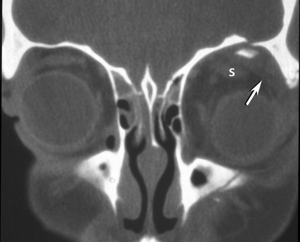
Angulated displaced fractures fragments are seen projecting downwards within the orbit indenting the superior rectus muscle.

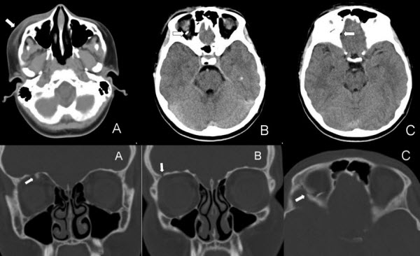
Orbital roof fracture radiology.

The orbital roof is composed of the orbital plate of the frontal bone with a small contribution from the lesser wing of the sphenoid at the apex figures 3 4 and 3 5. Fracture of the left zygomaticomaxillary attachmnet with fracture of the anterior and lateral maxillary walls inferior orbital rim fracture and left maxillary hemosinus. Dr robert foley and assoc prof frank gaillard et al. Very rarely the orbital roof will fracture without displacement of fractures fragments resulting in the non displaced orbital roof fracture.

Assessing traumatic orbital injuries is an important challenge for radiologists. Isolated orbital roof fractures in adults are uncommon comprising 12 19 of all orbital wall fractures. Ct scan revealed widening of the frontal bone fracture and the left orbital roof fracture with a hypodense cystic swelling in the left orbit and upper eyelid and left frontal lobe contusion. This is typically caused by a direct blow to the central orbit from a fist or ball.

Displaced orbital roof fracture 3. Although common in the pediatric population isolated orbital roof fractures can also occasionally occur in an adult fig 13. Most roof fractures are associated with other orbital fractures and result from significant head trauma as a high degree of force is required to fracture this portion of the orbit. A orbital fractures can occur in isolation or with other fracture patterns.

Radiological considerations in the setting of orbital roof fracture it is important to fully assess the extent of the fracture including any radiographically apparent concomitant abnormalities. As an isolated orbital roof fracture is a rare entity it is important to. Orbital fracture pearls are as follows. Orbital roof fracture icd 801 01 etiology.

This assessment is even more difficult when the orbital injury is associated with injuries involving multiple organs. A three dimensional ct reconstruction demonstrated the extent and shape of the skull fracture fig. The injury can be associated with a violation of the dura necessitating an intracranial approach. Orbital blowout fractures occur when there is a fracture of one of the walls of orbit but the orbital rim remains intact.

Communited mildly depressed left orbital roof fracture. Radiological considerations in the setting of orbital roof fracture it is important to fully assess the extent of the fracture including any radiographically apparent concomitant abnormalities.

Orbital Roof Blow In Fracture A Case Report And Review Jones Journal Of Radiology Case Reports

Orbital Roof Blow In Fracture A Case Report And Review Jones Journal Of Radiology Case Reports

Blow Out Fracture The Most Common Portion Of The Orbit To Sustain A Fracture Is The Weak Floor And This Injury If Occurring In Isolation May Result In A Blow

Blow Out Fracture The Most Common Portion Of The Orbit To Sustain A Fracture Is The Weak Floor And This Injury If Occurring In Isolation May Result In A Blow

Teardrop Sign Of Orbital Blowout Fracture Radiology Case Radiopaedia Org

Teardrop Sign Of Orbital Blowout Fracture Radiology Case Radiopaedia Org

Pin On Radiology Spotters

Pin On Radiology Spotters

Orbital Fracture Radiology Case Radiopaedia Org

Orbital Fracture Radiology Case Radiopaedia Org

Pathological Clavicle Fracture Radiology Case Radiopaedia Org Radiology Pathology Clavicle

Pathological Clavicle Fracture Radiology Case Radiopaedia Org Radiology Pathology Clavicle

Traumatic Orbital And Occular Injury Radiology Key

Traumatic Orbital And Occular Injury Radiology Key

Blowout Orbital Fracture Radiology Medical Imaging Radiography

Blowout Orbital Fracture Radiology Medical Imaging Radiography

Orbital Roof Fractures A Clinically Based Classification And Treatment Algorithm Omfs

Orbital Roof Fractures A Clinically Based Classification And Treatment Algorithm Omfs

Osteoma Of Orbital Roof Radiology Case Radiopaedia Org Radiology Radiology Imaging Diagnostic Imaging

Osteoma Of Orbital Roof Radiology Case Radiopaedia Org Radiology Radiology Imaging Diagnostic Imaging

Radial Head Fracture Radiology Case Radiopaedia Org Radiology Fractures Medical School Stuff

Radial Head Fracture Radiology Case Radiopaedia Org Radiology Fractures Medical School Stuff

Facial Bone X Ray Lateral View Www Anatomynote Com Facial Bones Radiology Radiology Humor

Facial Bone X Ray Lateral View Www Anatomynote Com Facial Bones Radiology Radiology Humor

Infantile Head Trauma With Orbital Roof Fracture Radiology Case Radiopaedia Org

Infantile Head Trauma With Orbital Roof Fracture Radiology Case Radiopaedia Org

Pxa Or Extra Ventricular Supratentorial Ependymomoa Which Is Common To Show Cyst And Nodule Cysts Personalized Items Head And Neck

Pxa Or Extra Ventricular Supratentorial Ependymomoa Which Is Common To Show Cyst And Nodule Cysts Personalized Items Head And Neck

Multiple Myeloma Is The Most Common Primary Malignant Bone Neoplasm In Adults And Results In A Wide Range Of Ra Radiology Multiple Myeloma Pediatric Radiology

Multiple Myeloma Is The Most Common Primary Malignant Bone Neoplasm In Adults And Results In A Wide Range Of Ra Radiology Multiple Myeloma Pediatric Radiology

International Day Of Radiology Radiopaedia Org Radiology Progressive Supranuclear Palsy Radiology Imaging

International Day Of Radiology Radiopaedia Org Radiology Progressive Supranuclear Palsy Radiology Imaging

Mount Fuji Sign Is Seen On Cross Sectional Imaging And Implies Tension Pneumocephalus Is Present The Sign Refers To The Pre Radiology Sinusitis Brain Images

Mount Fuji Sign Is Seen On Cross Sectional Imaging And Implies Tension Pneumocephalus Is Present The Sign Refers To The Pre Radiology Sinusitis Brain Images

Vocal Cord Paralysis Radiology Reference Article Radiopaedia Org In 2020 Thyroid Surgery Paralysis Nerve Palsy

Vocal Cord Paralysis Radiology Reference Article Radiopaedia Org In 2020 Thyroid Surgery Paralysis Nerve Palsy

1

1

Figure 6 From Orbital Fractures Role Of Imaging Semantic Scholar

Figure 6 From Orbital Fractures Role Of Imaging Semantic Scholar

Plasmacytoma Radiology Case Radiopaedia Org Radiology Case Limb

Plasmacytoma Radiology Case Radiopaedia Org Radiology Case Limb

Renal Cell Carcinoma Radiology Case Radiopaedia Org Renal Cell Carcinoma Radiology Radiology Imaging

Renal Cell Carcinoma Radiology Case Radiopaedia Org Renal Cell Carcinoma Radiology Radiology Imaging

Pin On Surgery

Pin On Surgery

Frontal Bone Fracture With Subdural Hematoma Radiology Case Radiopaedia Org

Frontal Bone Fracture With Subdural Hematoma Radiology Case Radiopaedia Org

Source : pinterest.com