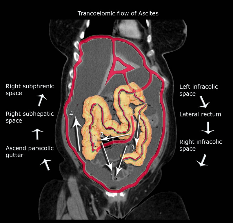
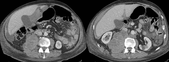
Paracolic Gutter Ascites
Epos Trade
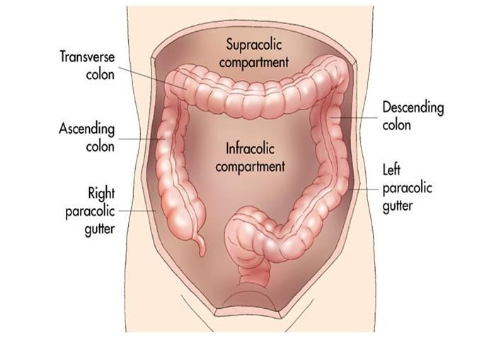
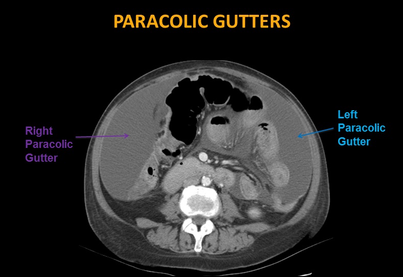
Paracolic Gutter
Epos

What Are The Paracolic Gutters Youtube

The Axial Ct Scan Shows Tumorous Masses Occupying The Right Lateral Download Scientific Diagram
Abdominal Xray Ct Us Radiology Flashcards Memorang

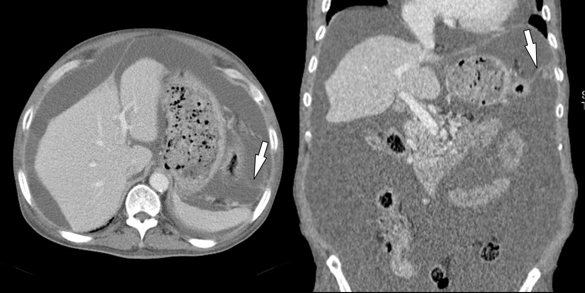
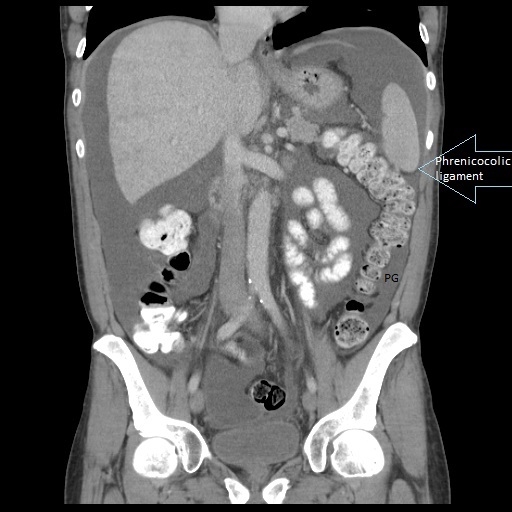
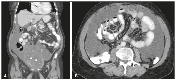
Visualized pancreas shows dilated main pancreatic duct 4 5 mm.
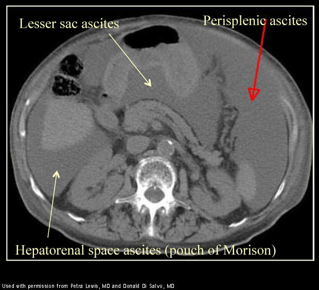
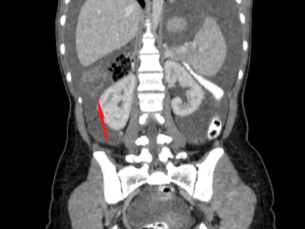
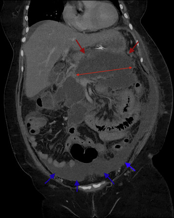
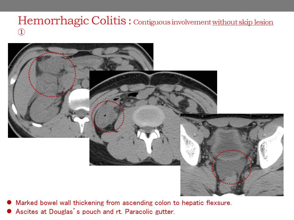
Paracolic gutter ascites.

The Abdominal Wall And Peritoneal Cavity Radiology Key
Epos Trade

Pancreatic Ascites Radiology Case Radiopaedia Org
Epos Trade

Fluid In The Paracolic Gutter Youtube
Chylous Ascites An Unusual Complication Of Necrotizing Pancreatitis The American Journal Of Medicine Blog
Abdomen Nontraumatic Emergencies Radiology Key
Epos Trade
Epos Trade
Https Www Abdominalradiology Org Resource Resmgr Eduposters 2016posters 29 Huang Pdf
The Radiology Assistant Peritoneal Pathology
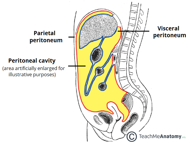
The Peritoneal Cavity Greater Sac Lesser Sac Teachmeanatomy
Http Pdf Posterng Netkey At Download Index Php Module Get Pdf By Id Poster Id 115818
Https Www Ajronline Org Doi Pdf 10 2214 Ajr 126 5 974
Https Onlinelibrary Wiley Com Doi Pdf 10 7863 Jum 1984 3 4 169

Abdomen Forum Neuronal Cell Body For Preganglionic Sympathetic Innervation To Stomach Iml 2 Neuronal Cell Body For Gva From Descending Colon Ppt Video Online Download

Epos Trade
Epos Trade
Https Encrypted Tbn0 Gstatic Com Images Q Tbn 3aand9gcshgccvhjwl4fga6xmtvrtmvxwjd3da6xh699xbs0yifrpvajgq Usqp Cau
Radiologia Brasileira As Multiplas Faces Do Pseudomixoma Peritonial Uma Revisao Radiologica Baseada Em 30 Casos
Http Journals Sagepub Com Doi Pdf 10 1177 87579302018003013

Ascites And Peritoneal Fluid Collections Radiology Key
Epos
Epos
Source : pinterest.com
