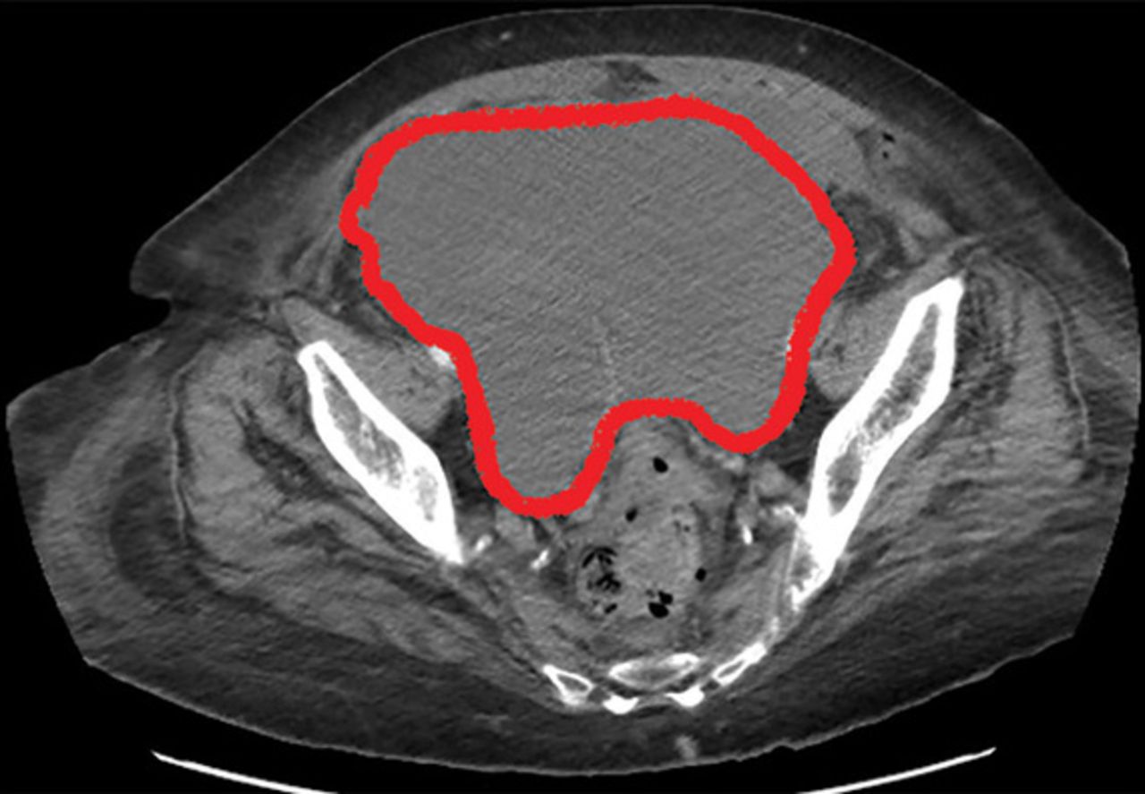
Paracolic Gutter Hematoma

Laparoscopic View Of Left Paracolic Gutter Hematoma Download Scientific Diagram
A Complication Of Enoxaparin Injection Cleveland Clinic Journal Of Medicine

Subcapsular Haematoma Of The Spleen Complicating Acute Pancreatitis Bmj Case Reports
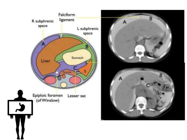
Peritoneum Intraperitoneal Spaces

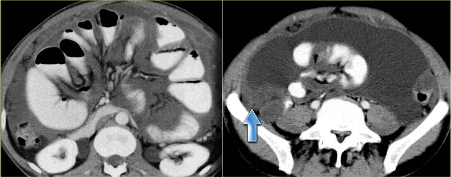
Ultrasonogram Revealed Free Fluid In The Paracolic Gutter Right And Download Scientific Diagram

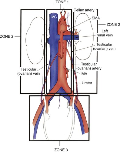
Amicus Illustration Of Amicus Injury Abdominal Abdomen Intraperitoneal Fluid Pericolic Gutter Dilation Small Bowel Side Wall Pelvic Hematoma Increased Blood Extraperitoneal Spaces Retrovessical Pouch Bladder Compressed Perisplenic Fracture Laverations
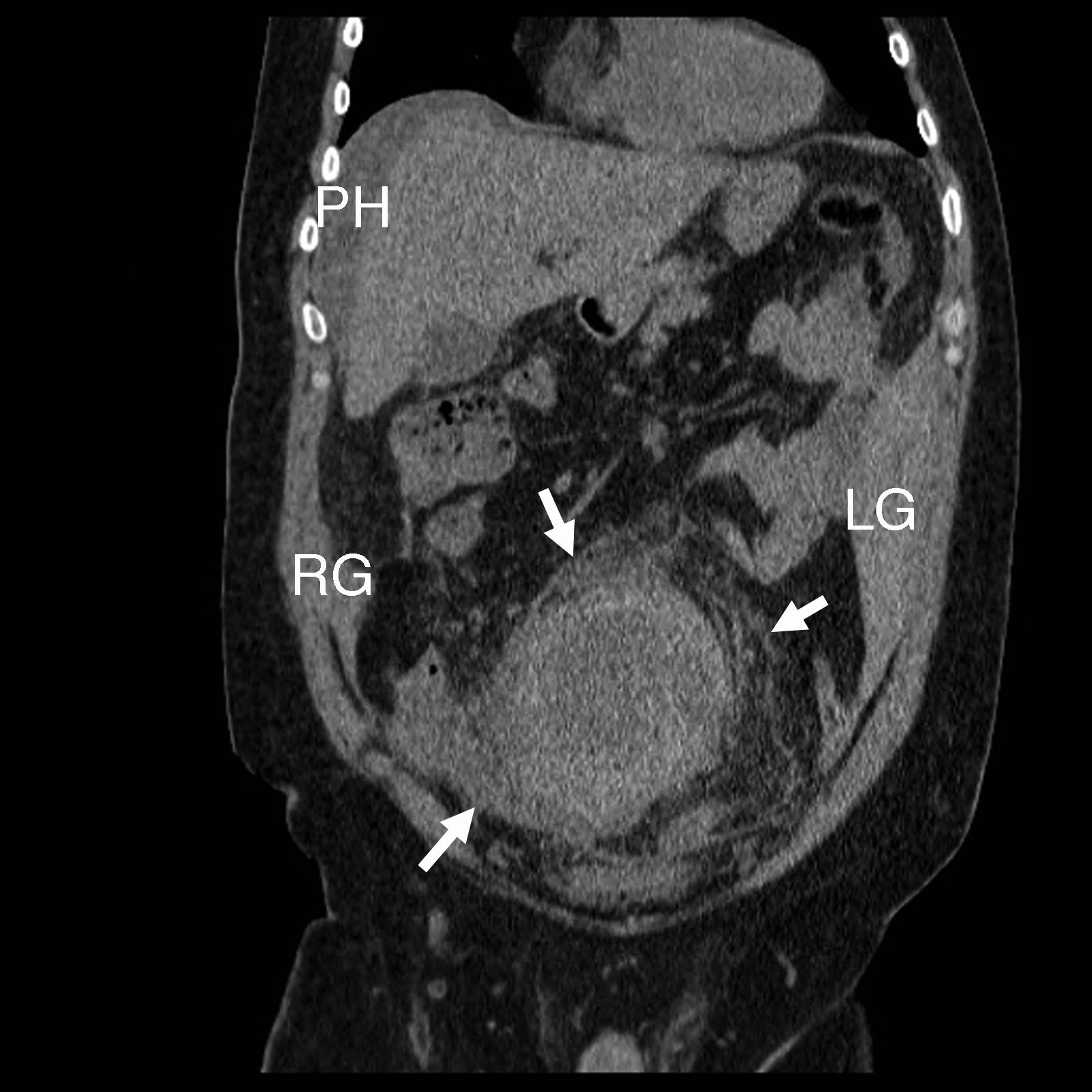
Spontaneous abdominal hemorrhage is defined as the presence of intraabdominal hemorrhage from a nontraumatic and noniatrogenic cause.
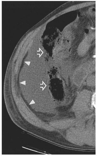
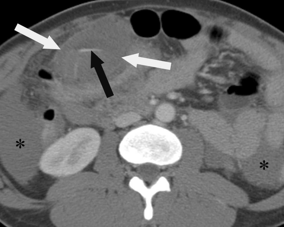
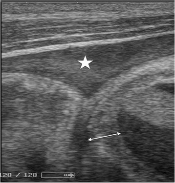
Paracolic gutter hematoma.

Ultrasonogram Revealed Free Fluid In The Paracolic Gutter Right And Download Scientific Diagram
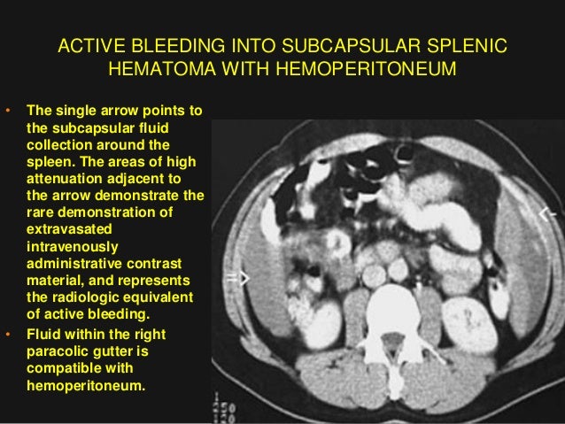
Imaging Abdomen Trauma Spleenic Trauma Part 3 Dr Ahmed Esawy

Ultrasound Image Of A Hemoperitoneum Star In The Right Paracolic Download Scientific Diagram
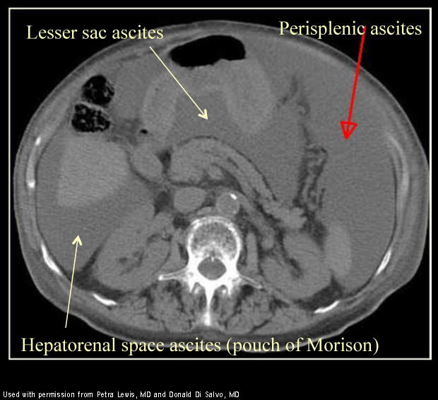
The Radiology Assistant Peritoneal Pathology
Plos One Percutaneous Image Guided Biopsy For Non Mass Forming Isolated Splenomegaly And Suspected Malignant Lymphoma

There Was A Massive Hematoma In The Peritoneum Surrounding The Spleen Download Scientific Diagram
Epos Trade

Figures Figure 1 Further Investigation With A Ct Abdomen And Pelvis Download Scientific Diagram

Figure 2 From A Case Of Retroperitoneal Ectopic Pregnancy Semantic Scholar

Fluid In Both Paracolic Gutters 33 Year Old Woman Involved In Metor Download Scientific Diagram
Abdomen Nontraumatic Emergencies Radiology Key
Epos Trade
Http Pdf Posterng Netkey At Download Index Php Module Get Pdf By Id Poster Id 115818
Abdominal Xray Ct Us Radiology Flashcards Memorang

Widespread Contrast Media Leak Within The Peritoneal Cavity Black Download Scientific Diagram

The Spread And Localization Of Intraperitoneal Abscesses Dynamic Radiology

Positive Right Upper Quadrant Ruq Fast View Showing Superior Download Scientific Diagram

Peritoneal Cavity And Abdominal Wall Radiology Key
Https Encrypted Tbn0 Gstatic Com Images Q Tbn 3aand9gcqfvuhmwj J7lodkwz Xnwkogimh7rxbftw9gin3yk76idtaqmi Usqp Cau

Gastrointestinal Radiology

Ct Scan Intraparenchymal Hepatic Hematoma With Active Bleeding From Download Scientific Diagram
Contribution Of Ultrasonography To The Diagnosis Of Internal Bleeding In Snakebite Envenomation Journal Of Venomous Animals And Toxins Including Tropical Diseases Full Text
Vascular Trauma Thoracic Key

Bowel Wall Thickening In Children Ct Findings Ppt Download
Source : pinterest.com
